Featured Course
Introduction to Vascular Ultrasound
This course is designed to provide an introduction into the ultrasound evaluation of the cerebrovascular system, lower extremity arterial system and lower extremity venous system.
VIEW TRAINING COURSE
Case Study Details
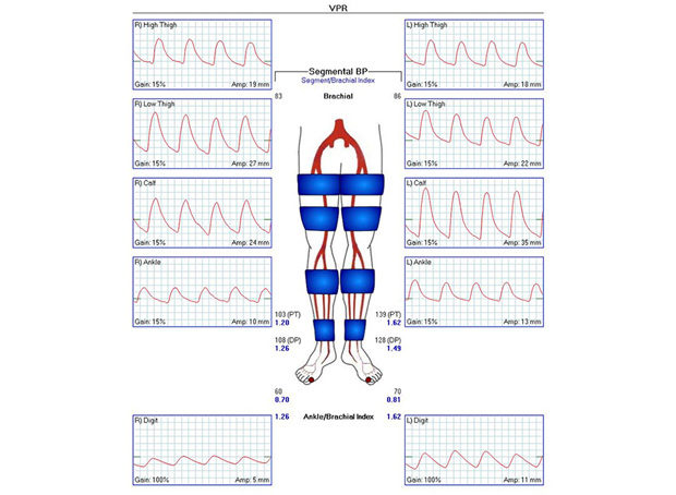
While auscultating Doppler signals at the wrists for blood pressure measurement, the sonographer noted that the Doppler signals were audibly monophasic. Brachial pressures measured 83 mm Hg on the right and 86 mm Hg on the left. Because of the low arm pressures and monophasic Doppler signals, the sonographer decided to check the subclavian arteries using duplex ultrasound.
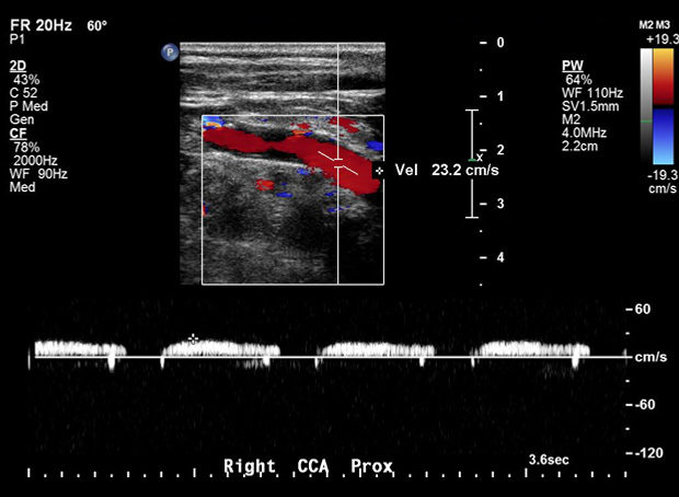
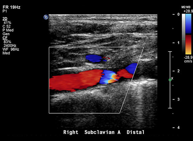
On the right, innominate, proximal to mid subclavian, and vertebral artery flow was retrograde. Distal subclavian artery flow was antegrade, with a collateral vessel noted at the mid to distal subclavian artery. Common carotid artery flow was very low and monophasic. Findings suggest significant proximal innominate artery obstruction.
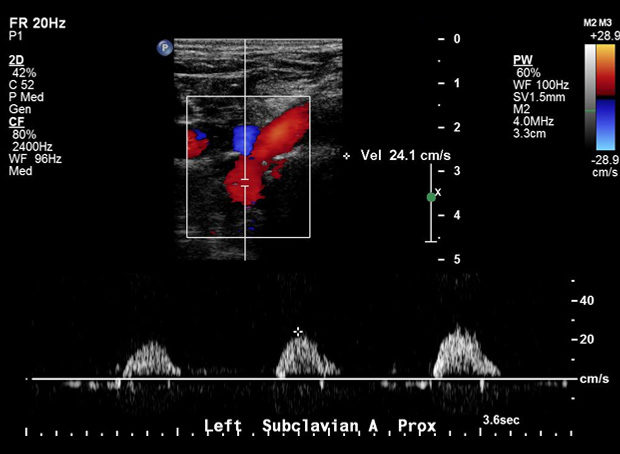
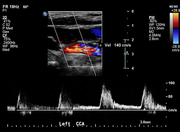
On the left, subclavian and vertebral artery flow was antegrade but monophasic. Common carotid artery flow was essentially normal. Findings suggest proximal left subclavian artery obstruction.
Carotid Duplex revealed plaque at the carotid bifurcations bilaterally, however velocity criteria is not applicable due to proximal obstruction. Conventional Angiogram shows: Innominate and Left prox SCA occlusion. LICA 40%. R carotid, SCA & LSCA fill via
collaterals from L carotid system via vertebrals, RICA. LSCA stented.”
Tell Us Your Story
We want to hear how you are using Ultrasound to improve patient care. Email Us Your Story