Featured Course
Vascular Interpretation Preceptorship
This Vascular Interpretation Preceptorship combines up to 3 days of in-person lecture, discussion and collaborative learning, with access to our library of over 500 vascular cases for interpretation and assessment. VIEW TRAINING COURSE
Case Study Details
A 49 year old Hispanic female.
History of stroke two years ago with residual dysarthria and right hemiparesis.
Carotid Duplex
The patient came to her appointment at the Vascular Lab as part of a standard pre-operative evaluation.
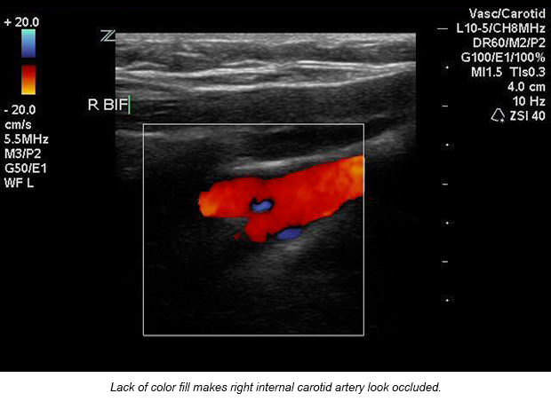
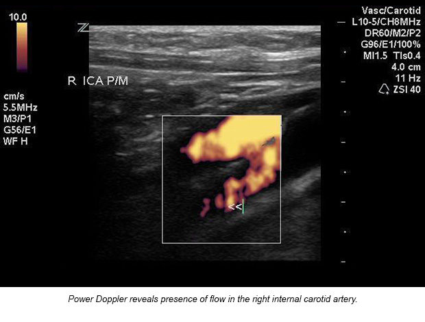
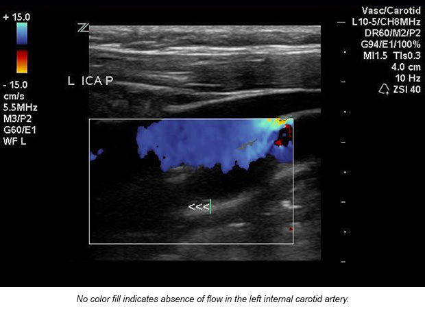
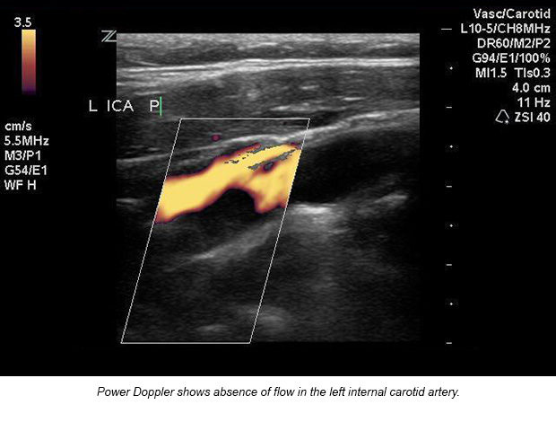
Utilizing power Doppler to discern occlusion from near-occlusion.
Ultrasound diagnosis: 70-99% right internal carotid artery stenosis and left internal carotid artery occlusion. Surgery cannot be performed on an occluded internal carotid artery. Correlating with the ultrasound, CT diagnosed Rose with a 99% right internal carotid artery stenosis and 100% left internal carotid artery occlusion. CT of her brain showed only an old left frontal lobe infarct, consistent with her right hemiparesis.
Due to the location of the right internal carotid artery stenosis in the bony canal and full occlusion of the left internal carotid artery, the physician concluded that carotid surgery was not an option for the patient, but expressed that she is likely to continue functioning due to collateral flow around her carotid arteries.
Tell Us Your Story
We want to hear how you are using Ultrasound to improve patient care. Email Us Your Story


